Navigating the Narrowing Path: A Comprehensive Guide to Spinal Stenosis
Our spinal cord and nerves must travel a path. Narrowing of the path can cause pain- and much worse. Loss of strength, balance, sensation, or even loss of bowel and bladder control can be the result of spinal stenosis. And time is not on your side. Longer term compression can lead to nerve damage that doesn’t recover.
Spinal stenosis is a common and often debilitating condition that affects millions, leading to persistent back pain, neck pain, and radiating symptoms like sciatica. If you’re experiencing these issues, understanding what spinal stenosis is, what causes it, and the range of treatment options available, including conservative care like chiropractic, is crucial. This article will serve as your guide to this complex spinal condition, helping you make informed decisions about your health.
What is Spinal Stenosis?
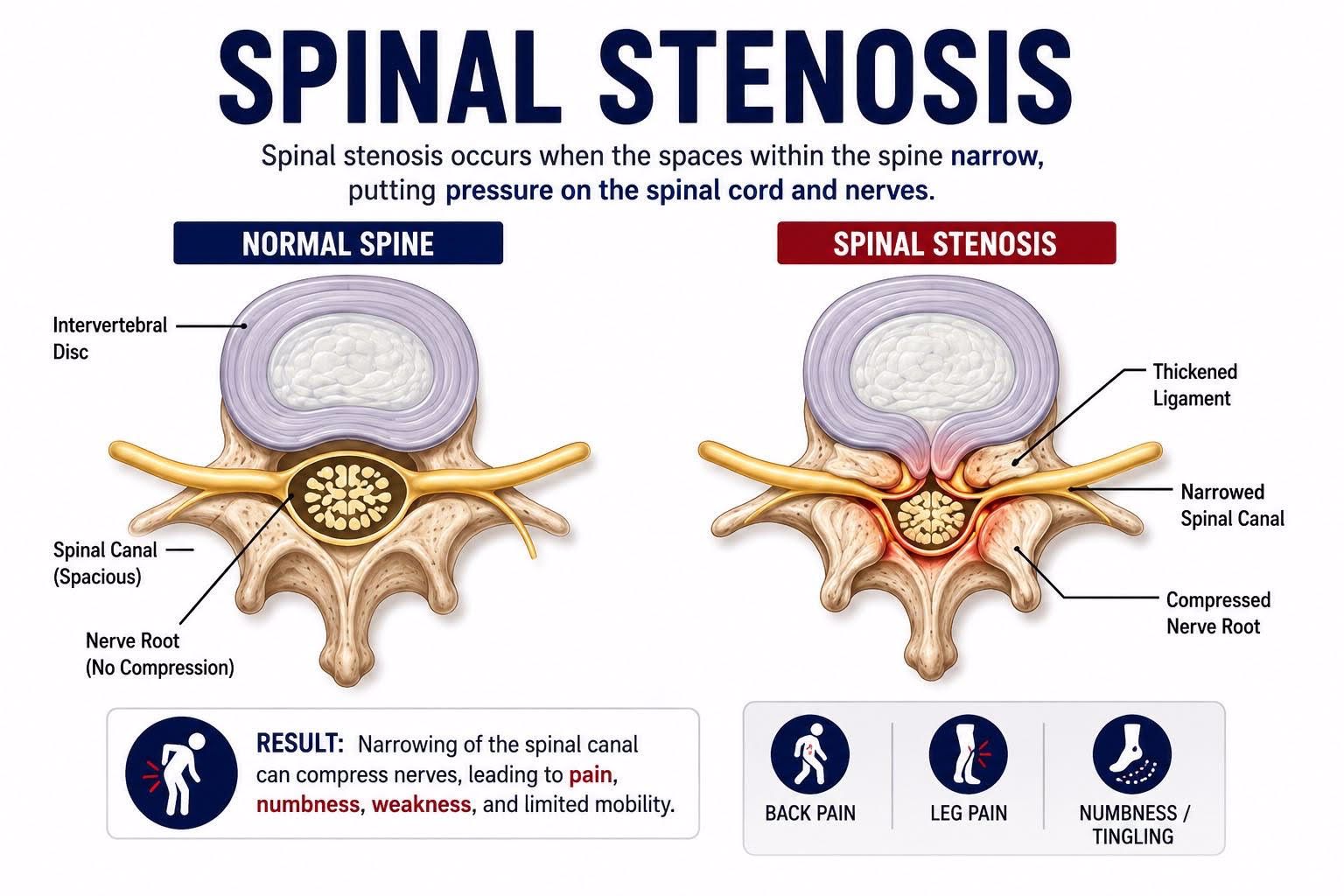
At its core, spinal stenosis refers to the narrowing of the spaces within your spine, which can put pressure on the spinal cord and the nerves that branch off it. This narrowing can occur in the cervical spine (neck), leading to cervical spinal stenosis, or more commonly, in the lumbar spine (lower back), resulting in lumbar spinal stenosis.
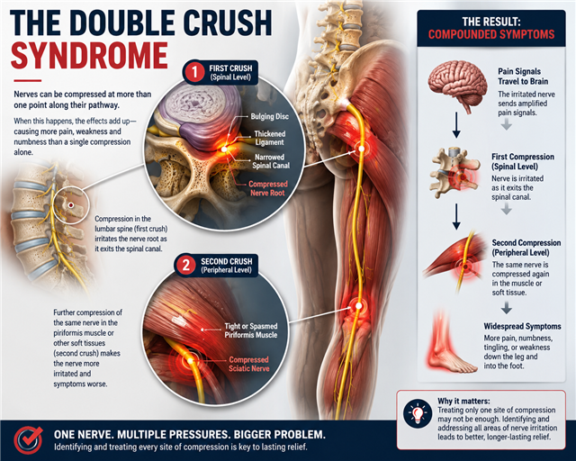
The spinal stenosis symptoms you experience can be incredibly varied and are often multifactorial. While direct compression of nerves within the spine is a primary culprit, it’s essential to consider the possibility of peripheral entrapments. This means that nerves can be compressed at multiple points along their pathway, not just within the spinal canal. For instance, someone with lumbar spinal stenosis might also have a peripheral nerve entrapment in the piriformis muscle, contributing to their sciatica and leg pain. This interplay of multiple entrapments can complicate symptom presentation and diagnosis.
The “stenosing” itself isn’t a single entity. It can involve several anatomical changes:
• Bony Stenosing: This often results from the growth of bone spurs (osteophytes) on the vertebrae, a natural part of the degenerative process. These bony growths can intrude into the spinal canal or the neuroforamina (the openings where nerves exit the spine).
• Disc Tissue Involvement: Degenerative disc disease is a major contributor. As intervertebral discs dehydrate and lose height, they can bulge or herniate into the spinal canal. A herniated disc can directly compress nerves, exacerbating the narrowing.
• Ligamentum Infolding: The ligaments that support the spine, particularly the ligamentum flavum, can thicken and buckle into the spinal canal as they degenerate. This infolding significantly reduces the available space for the spinal cord and nerves.
• Alignment Qualification: Changes in spinal alignment, such as spondylolisthesis (where one vertebra slips forward over another) or exaggerated spinal curves, can also contribute to spinal stenosis by altering the dimensions of the spinal canal and putting additional stress on surrounding structures.
Understanding these various contributing factors is key to appreciating the complexity of spinal stenosis pain and why a comprehensive approach to diagnosis and treatment is necessary.
What Causes Spinal Stenosis?
Spinal stenosis is predominantly a degenerative spine condition, meaning it develops gradually over time due to wear and tear on the spine. Age is the most significant risk factor, as the natural aging process leads to changes in the bones, discs, and ligaments of the spine.
Common causes include:
• Osteoarthritis: The most frequent cause, leading to bone spur formation and ligament thickening.
• Degenerative Disc Disease: As discs lose water content and height, they can bulge or herniate, narrowing the spinal canal.
• Thickening of Ligaments: The ligamentum flavum, a key ligament supporting the spine, can thicken and buckle into the spinal canal.
• Spondylolisthesis: The slipping of one vertebra over another can significantly reduce the space for nerves.
• Spinal Tumors: Though less common, tumors can grow within the spinal canal, causing compression.
• Spinal Injuries: Trauma can lead to fractures or dislocations that narrow the spinal canal.
• Postural Distortions: This element of stenosis often escapes traditional testing.
A crucial concept in understanding nerve compression, especially in spinal stenosis, is the double-crush phenomenon. This speaks to the effect of a summation of multiple entrapens along a neuron pathway on the same nerve, a nerve that has been compressed or irritated at one site becomes more susceptible to damage from compression at another site along its pathway. For example, a nerve root compressed by a bulging disc in the lumbar spine (the “first crush”) might then be more susceptible to being compressed by tight muscles or fascia in the buttock or leg (the “second crush”). This summation of multiple nerve entrapments can lead to more severe and widespread symptoms than either compression alone, explaining why some individuals experience significant leg weakness or numbness in the feet even with seemingly moderate spinal stenosis. This concept underscores the importance of a thorough examination that considers the entire nerve pathway.
Diagnostic Testing for Spinal Stenosis
Accurate diagnosis is paramount for effective spinal stenosis treatment. A comprehensive evaluation typically involves:
•Detailed Medical History and Physical Examination: Your healthcare provider will ask about your symptoms, their duration, what makes them better or worse, and your overall health. A physical exam will assess your range of motion, muscle strength, reflexes, and sensory function to identify patterns of nerve compression.
•X-rays: Can reveal bony changes, such as bone spurs, loss of disc height, and spinal alignment issues like spondylolisthesis. While X-rays don’t show soft tissues, they are a good starting point.
•Magnetic Resonance Imaging (MRI): This is considered the gold standard for diagnosing spinal stenosis because it provides detailed images of soft tissues, including discs, ligaments, spinal cord, and nerve roots. MRI can identify areas of narrowing caused by bony stenosis, disc bulging or herniation, ligament thickening, facet joint changes, and spinal alignment issues. It can show the location and severity of central canal, lateral recess, and foraminal stenosis. However, MRI findings must always be interpreted along with the patient’s symptoms and physical examination, as some people may have significant narrowing without symptoms, while others may experience symptoms with less obvious imaging findings.
•Computed Tomography (CT) Scan: Often used if MRI is contraindicated or to provide more detailed images of bone structures, especially after trauma or surgery. A CT myelogram, which involves injecting dye into the spinal canal, can offer even clearer images of nerve compression.
“How is spinal stenosis diagnosed?” It’s a combination of your symptoms, a thorough physical exam, and specific imaging studies that paint a clear picture of the underlying anatomical changes.
Conservative Care vs. Surgery for Spinal Stenosis
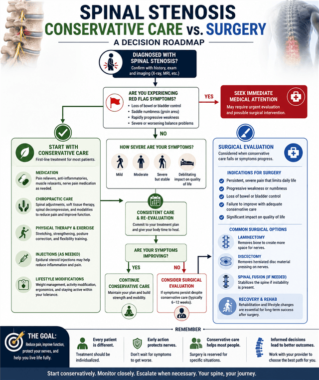
When facing a diagnosis of spinal stenosis, a common question is, “Do I need surgery for spinal stenosis?” For the vast majority of individuals, non-surgical spinal stenosis treatment is the first and often most effective course of action. The care provider and the patient must be mindful of the dangers of prolonged stenosing and the signs of nerve damage. Treating any nerve entrapment, to include stenosing, means to reduce or alleviate the entrapment. Caring for the nerve affectation (therapeutically as well as nutritionally) should also be considered. There could be a scenario where delaying indicated surgery is counterproductive. Conservative care that isn’t showing demonstrable signs of success should not be prolonged.
Conservative Care for Spinal Stenosis
Conservative treatments aim to manage spinal stenosis symptoms, improve function, and reduce stress on irritated spinal structures. These approaches may include:
• Medication: Over-the-counter pain relievers, anti-inflammatory medications, muscle relaxants, and, in some cases, medications designed to help manage nerve-related pain.
•Physical Therapy: A structured exercise program can help improve strength, flexibility, posture, balance, and walking tolerance. Therapists may also use techniques such as heat, ice, and electrical stimulation to help manage symptoms.
•Injections: Epidural steroid injections may provide temporary relief by reducing inflammation around irritated nerve roots. These are typically used as part of a broader treatment plan rather than a standalone solution.
•Chiropractic Care: Chiropractic care provides a conservative approach focused on improving spinal mobility, reducing mechanical stress, addressing contributing biomechanical factors, and helping patients maintain function. Treatment is individualized and may include spinal adjustments, soft tissue techniques, therapeutic exercises, and other supportive therapies.
•Lifestyle Modifications: Maintaining a healthy weight, improving posture, modifying aggravating activities, and staying physically active can help reduce symptom flare-ups and support long-term spinal health. For many people with lumbar spinal stenosis, walking can be beneficial when performed within their tolerance level. Claudication-increased stenosis symptoms with prolonged walking-may require further limit on walking. Some individuals may need to adjust walking duration, pace, or terrain based on their symptoms.
When Is Surgery Necessary for Spinal Stenosis?
Surgery is generally considered when conservative treatments have failed to provide adequate relief, or when there are signs of progressive neurological deficits such as:
• Significant or worsening leg weakness.
• Loss of bowel or bladder control (cauda equina syndrome, a medical emergency).
• Intractable pain that severely impacts quality of life.
The most common surgical procedures for spinal stenosis include:
• Laminectomy: Removal of part of the vertebral bone (lamina) to create more space for the spinal cord and nerves.
• Discectomy: Removal of a herniated disc fragment.
• Spinal Fusion: In some cases, if instability is present, vertebrae may be fused together after decompression.
While surgery can be effective, it carries risks and requires a significant recovery period. Many people find excellent relief through conservative measures, making it the preferred initial strategy.
Red Flags: When Spinal Stenosis Requires Immediate Medical Attention
Although spinal stenosis usually develops gradually and can often be managed successfully with conservative care, certain symptoms may indicate significant nerve compression or spinal cord involvement. These “red flag” symptoms should never be ignored and warrant prompt medical evaluation.Seek Immediate Medical Care If You Experience:
• Loss of bowel or bladder control. Difficulty controlling urination or bowel movements, or being unable to empty your bladder, may indicate cauda equina syndrome, a rare but serious condition requiring emergency surgical evaluation.
• Numbness in the groin or saddle region. Loss of sensation in the inner thighs, buttocks, or genital area (“saddle anesthesia”) is another warning sign of possible cauda equina syndrome.
• Rapidly progressing leg weakness. Increasing difficulty lifting your foot, climbing stairs, standing from a seated position, or walking may indicate worsening nerve dysfunction. Progressive weakness should be evaluated promptly to reduce the risk of permanent nerve damage.
• Significant loss of balance or coordination. In cervical spinal stenosis, compression of the spinal cord (cervical myelopathy) can cause problems with balance, hand coordination, fine motor skills, or frequent falls. These symptoms require timely assessment by a spine specialist.
• Severe pain following trauma. New or worsening neck or back pain after a fall, motor vehicle accident, or other significant injury should be evaluated to rule out fractures or spinal instability.
• Persistent numbness that continues to worsen. While intermittent tingling or numbness is common with spinal stenosis, symptoms that steadily progress or spread to new areas can indicate the need for escalated care.
Chiropractic Treatment for Spinal Stenosis
“Can chiropractic help spinal stenosis?” Absolutely. Chiropractic care offers a holistic, non-surgical approach that focuses on improving spinal function, reducing nerve irritation, and enhancing the body’s natural healing capabilities. A chiropractor for spinal stenosis in Augusta (or wherever you are located) is a spine specialist who can develop a personalized chiropractic treatment for spinal stenosis plan.
Chiropractic treatment for spinal stenosis typically includes:
• Spinal Adjustments (Manipulations): Gentle, specific adjustments are used to restore proper motion to restricted spinal joints. While chiropractic adjustments don’t “widen” the spinal canal, they can improve the common aperture (formen by contiguous vertebrae) through which the cord must pass as well as improve biomechanics, reduce inflammation, and decrease muscle spasm, thereby alleviating pressure on nerves. “Can spinal adjustments help spinal stenosis?” Yes, by improving overall spinal mechanics and reducing compensatory stress.
• Electrical Stimulation: Helps reduce pain and muscle spasms.
• Heat and Ice Therapy: Applied to reduce inflammation and relax tight muscles.
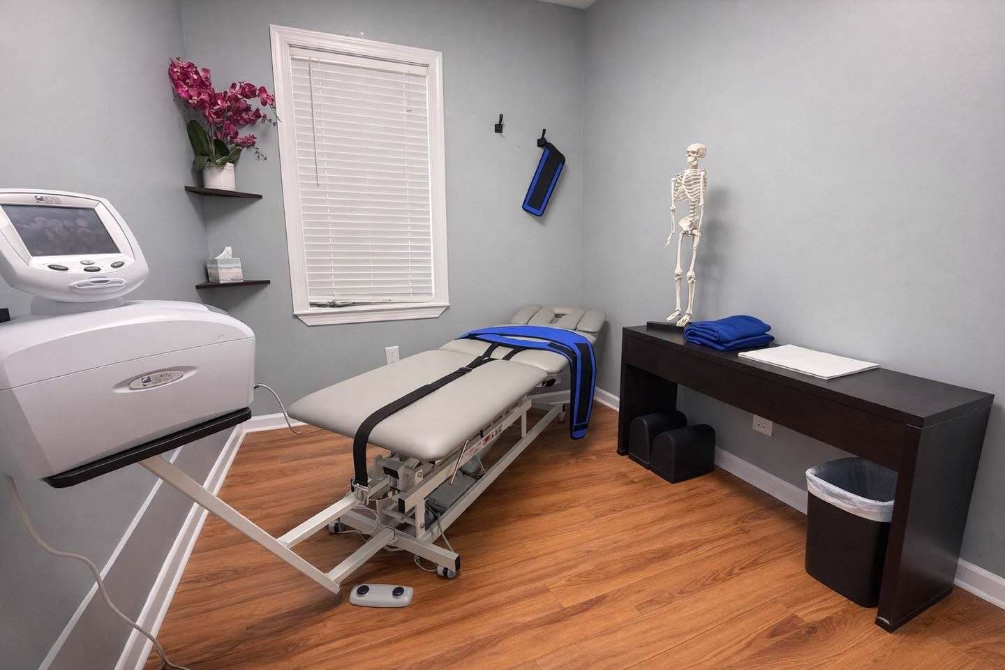
• Spinal Decompression for Spinal Stenosis: This is a highly effective non-surgical spine treatment. Spinal decompression therapy involves a specialized table that gently stretches the spine, creating negative pressure within the discs. This can help to:
• Create more space for compressed nerves.
• Reduce pressure on bulging or herniated discs.
• Promote the influx of nutrient-rich fluids into the disc space, aiding healing.
• Relieve pressure from facet joints and surrounding ligaments.
Many patients searching lumbar spinal stenosis treatment near me or cervical spinal stenosis chiropractor seek out decompression as a primary treatment option.
• Active Rehabilitation and Therapeutic Exercises: A crucial component of long-term success. Your chiropractor will prescribe specific exercises tailored to your condition to:
• Strengthen core muscles, improving spinal stability.
• Increase flexibility and range of motion.
• Improve posture and body mechanics.
• Examples of best exercises for spinal stenosis often include flexion-based exercises that help to open the spinal canal, such as pelvic tilts, knee-to-chest stretches, and gentle abdominal strengthening. Education on proper body mechanics for daily activities is also vital.
• Lifestyle and Ergonomic Counseling: Advice on diet, hydration, posture, and activity modification to support spinal health and prevent symptom exacerbation.
The goal of chiropractic care is not just pain relief but also to address the underlying biomechanical dysfunctions contributing to the stenosis, helping you to “get better without surgery” and manage your condition effectively. For those seeking natural treatment for spinal stenosis or comparing physical therapy vs chiropractic for spinal stenosis, it’s important to recognize that both can be highly beneficial and often complementary, with chiropractic emphasizing spinal alignment and nervous system function.
Taking Control of Your Spinal Stenosis
Living with spinal stenosis can be challenging, but it doesn’t have to define your life. Understanding what does spinal stenosis feel like – often a dull ache, radiating pain, numbness, or weakness that worsens with standing or walking and improves with sitting – is the first step toward seeking help. With a clear diagnosis and a commitment to conservative care, particularly chiropractic treatment for spinal stenosis, many individuals experience significant relief and an improved quality of life.
If you’re in the CSRA area and experiencing lower back pain, neck pain, or sciatica consistent with spinal stenosis, don’t hesitate to seek professional help. A qualified chiropractor for spinal stenosis in Augusta can offer personalized care, including spinal decompression, to help you navigate this narrowing path and find lasting relief.
References:
1. Deyo, R. A., & Mirza, S. K. (2016). Clinical practice. Lumbar spinal stenosis. New England Journal of Medicine, 374(25), 2419-2429.
2. Genevay, S., & Atlas, S. J. (2010). Lumbar spinal stenosis. Best Practice & Research Clinical Rheumatology, 24(2), 253-265.
3. Kreiner, D. S., et al. (2019). An evidence-based clinical guideline for the diagnosis and treatment of degenerative lumbar spinal stenosis. The Spine Journal, 19(3), 395-414.
4. Murphy, D. R., & Hurwitz, E. L. (2015). A nonsurgical approach to the management of lumbar spinal stenosis: a prospective observational study. Journal of Chiropractic Medicine, 14(4), 213-220.
5. Norman, S., & Shifflett, K. (2018). The Double Crush Syndrome. Journal of Orthopaedic & Sports Physical Therapy, 48(10), 833-834. (Conceptual reference for double crush)
6. Pope, M. H., & Frymoyer, J. W. (1995). The Lumbar Spine: Clinical Biomechanics and Management. Mosby. (General biomechanical principles)
7. Schonstrom, N., & Lindahl, S. (1984). The narrow lumbar spinal canal and the “epidural sign”: a myelographic investigation. Acta Radiologica: Diagnosis, 25(6), 465-472. (Early research on anatomical narrowing)
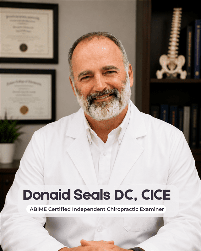
Written by: Donaid Seals D.C
Dr. Seals is a practicing Doctor of Chiropractic with over 25 years of experience caring for people. His thinking is the product of his education, practice experience and many years in the natural foods and fitness industry. He has become living proof that old muscleheads don’t die-or fade away; sometimes they grow up to bring real-world expertise to the clinical picture. Traditional background information is available here.
Disclaimer: Consult with a healthcare provider before starting any new supplement/workout regimen, especially if you have underlying health conditions or take medications. The entire contents of this website are based upon the opinions of Dr. Seals, unless otherwise noted. Individual articles are based upon the opinions of the respective author, who retains copyright as marked. The information on this website is not intended to replace a one-on-one relationship with a qualified health care professional and is not intended as medical advice. It is intended as a sharing of knowledge and information from the research and experience of Dr. Seals and his community. Dr. Seals encourages you to make your own health care decisions based upon your research and in partnership with a qualified health care professional.